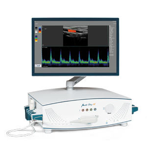
The Extracranial Doppler Sonography examines potential constrictions and occlusions of the carotid arteries. It is used for stroke prophylaxis and the clarification of circulatory disorders. The imaging method of the Extracranial Duplex Sonography, which is a combination of Doppler and Color Doppler Sonography, allows comprehensive statements regarding potential vascular changes. This method, which is also called Carotis Duplex Sonography, enables the display and measurement of the intima media thickness, makes changes of the vascular wall or of the vessel diameter visible and facilitates a prognostic assessment of the risks of cardiac and cerebral infarction, as well as of cardiovascular risks. Additionally, blood flows are made visible and blood flow velocities, which increase when vessels are constricted, are measured by means of the color-coded Doppler technology. In many cases, these increases are the first symptoms of an increased risk of stroke. This could be life-saving information to patients.
Applications of the Extracranial Color Doppler Sonography include:
- Aneurysms
- Arterial plaques
- Stenoses
- Vasospasms